A 3D echocardiogram in Dubai is one of the advanced non-invasive ways doctors can assess how your heart looks and works in real time. Unlike many tests that only provide static images, echocardiography uses ultrasound waves to create moving pictures of the heart, helping the cardiologist evaluate the heart chambers, valves, pumping function, and blood flow patterns. The American Heart Association explains that an echocardiogram uses sound waves to create pictures of the heart and provides details about heart structure and function.
At Gemelli Medical Centre in Jumeirah, cardiology care is positioned around non-invasive cardiovascular assessment, prevention, and risk stratification. The center’s cardiology page also highlights the use of Philips 3D Echo technology for detailed real-time imaging of the heart’s structure and function, especially in heart valve assessment and complex cardiac conditions.
Send us a Message
What Is a 3D Echocardiogram?
A 3D echocardiogram is a type of heart ultrasound that creates three-dimensional images of the heart. It allows the cardiologist to view the heart from multiple angles and better understand the shape, movement, and function of the heart valves and chambers.
A standard 2D echocardiogram is already useful for many routine assessments. However, a 3D echo can offer additional detail in selected cases, particularly when doctors need a clearer view of valve anatomy, heart chamber size, or structural abnormalities. The American Heart Association notes that 3D echocardiography may be used in diagnosing problems with the mitral valve.
When Might a Doctor Recommend an Echocardiography Test?
Your doctor may recommend an echocardiography test in Dubai if you have symptoms, risk factors, or clinical findings that require a closer look at the heart. Common reasons include:
- Chest discomfort or unexplained shortness of breath
- Palpitations or irregular heartbeat
- A heart murmur heard during examination
- High blood pressure affecting the heart
- Suspected valve narrowing or leakage
- Follow-up after a previous cardiac diagnosis
- Assessment of heart function before or after treatment
- Preventive evaluation in patients with risk factors
An echo can help doctors assess the size and shape of the heart, how well the heart pumps blood, whether the valves open and close properly, and whether there is valve leakage or narrowing.
How to Prepare for a Cardiac Ultrasound
For most transthoracic echocardiograms, preparation is simple. You can usually eat and drink normally unless your doctor gives different instructions. Wear comfortable clothing, and bring any previous cardiac reports, ECGs, blood tests, medication lists, or imaging results.
For better cardiac ultrasound preparation, patients should inform the medical team if they have:
- Previous heart surgery
- Known valve disease
- Pacemaker or implanted cardiac device
- Recent chest pain or fainting
- Current heart medications
- Pregnancy or relevant medical conditions
This information helps the cardiology team interpret the test in the correct clinical context.
What Happens During the Test?
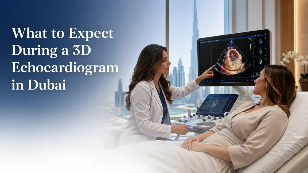
During a 3D echocardiogram, you will usually lie on an examination couch. A trained specialist applies ultrasound gel to the chest and uses a handheld probe called a transducer. The probe sends sound waves into the body and receives echoes that are converted into live heart images.
You may be asked to change position, hold your breath briefly, or lie on your left side. This helps obtain clearer images of the heart. The test is usually painless. You may feel light pressure from the probe on the chest, but it should not be uncomfortable.
Because echocardiography uses sound waves rather than ionizing radiation, it is generally considered a safe and non-invasive cardiac imaging test. The American Heart Association states that an echo does not hurt and has no side effects.
What Can a 3D Echo Show?
A 3D echo may help the cardiologist evaluate:
- Heart valve structure and motion
- Valve narrowing or leakage
- Pumping strength of the heart
- Chamber size and heart wall movement
- Possible causes of heart murmurs
- Signs of heart failure or cardiomyopathy
- Congenital or structural abnormalities
- Follow-up after certain cardiac procedures
This is why valve assessment in Dubai can benefit from advanced echocardiography. For example, in mitral valve problems, a 3D view may help the doctor understand the exact shape and behavior of the valve more clearly than 2D imaging alone.
When Will You Get the Results?
The timing depends on the clinic workflow and the complexity of the case. In many cases, the cardiologist or reporting specialist reviews the images and explains the key findings after assessment. At Gemelli Medical Centre, the care model emphasizes clear digital reports and structured explanations, helping patients understand the next steps rather than receiving technical results without context.
Your report may include information about:
- Ejection fraction or pumping function
- Valve function
- Chamber size
- Wall motion
- Pressure estimates
- Any abnormal findings requiring follow-up
Why Choose an Integrated Cardiology Setting?
A 3D echo is not only a “test”; it is part of a broader clinical decision. The value comes from combining your symptoms, medical history, physical examination, ECG, blood pressure, lab results, and imaging findings.
Gemelli Medical Centre describes its care journey as coordinated, where targeted evaluation, diagnostics, collaborative medical review, and clear next steps are part of the patient pathway. This supports stronger medical E-E-A-T because the test is interpreted inside a clinical framework, not as a standalone scan.
Final Takeaway
A 3D echocardiogram in Dubai can help patients and doctors understand heart structure and function in a detailed, non-invasive way. It is especially useful when assessing valves, heart function, murmurs, or symptoms such as breathlessness and palpitations.
If your doctor has recommended a heart ultrasound, or if you are concerned about your cardiac risk, booking a cardiology assessment can help determine whether a 2D echo, 3D echo, ECG, stress test, or other cardiac investigation is the right next step.
To discuss whether a 3D echocardiogram is suitable for your case, contact Gemelli Medical Centre in Jumeirah or book a cardiology assessment.
 English
  Arabic